Tissue processing
To make visible what we want to observe, it is necessary to implement various techniques that we apply to the material, this is called preparation of the samples. For observation in light microscopy or electron microscopy, the tissue sections examined are the result of technical procedures which require several successive steps: fixation, inclusion, cutting, staining, mounting.
The preparation of histological samples may vary greatly depending on the intrinsic properties of the samples such as size but also according to the objectives of the examination (histopathological diagnosis in humans or animals or research protocol) and procedures downstream of this preparation.
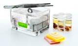
The purpose of fixation is to preserve the structures. Indeed, the tissue collection causes them to die: the cells discharge their enzymes, which causes a self-regulation of the tissue. Moreover, in the ambient air, samples can be contaminated with bacteria.
The purpose of inclusion is to allow fine and regular cuts. The most widely used inclusion medium is paraffin, but other media are used depending on the mode of preservation of the tissues and the techniques used.
The cuts of the paraffin block are made with a microtome allowing to make section slices fine enough to be observed by microscopy. The thickness of the sections depends on the microscopy technique used.
The stainings made on slides, accentuate the contrasts to better recognize the different elements of the preparation.
Finally the mounting is the last step before the observation.